Foods and phytonutrients that may benefit T cells

T cells are a group of white blood cells with immune functions that may be beneficial or which may promote inflammation. They can change from one type of T-cell to another based on the amount of oxidative stress chemicals present in their surroundings. Our own body can produce antioxidants that reduce oxidative stress when we have adequate Nrf2. We also can get antioxidants from foods and often the foods that are good sources of antioxidants also contain phytonutrients that promote our own production of Nrf2 which then can help us produce our own types of antioxidants. Other nutrients are also helpful for promoting T cells to become the less inflammatory producing types. (1)

Reduced antioxidant production and prevalence of the more inflammatory type of T cells may be involved in aging and many types of chronic illnesses including cardiovascular diseases, neurological disorders, carcinoma’s and leukemia’s, autoimmune disorders, inflammatory bowel disease and vitiligo. For more details see: (1) Figure 13)

See Table 3: Antioxidant Compounds of Natural Dietary Products with Role in T Cell Function. (1)

  • Green tea: Catechin hydrate (CH) and Epigallocatechin 3-gallate (EGCG). Teas (white, green, and oolong), cocoa, grapes, berries, apples; Catechins are monomers of Flavan-3-ols; see more:(Flavonoids)
  • Carrot, celeriac, parsnip, and parsley: Aliphatic C(17)-polyacetylenes. The Apiaceae plant family, see more: (Aliphatic C(17)-polyacetylenes) Commonly used herbs spices in the plant family include caraway seeds, coriander seeds/cilantro leaves, cumin seeds, dill, fennel; see more: (Apiaceae plants)
  • Turmeric (yellow spice in curry powder): Curcumin (diferuloylmethan). See more: (Curcumin)
  • Garlic: Ajoene. Organosulfur compound, others also present in onions, see more: (Ajoene/organosulfur compounds).
  • Plant foods/beverages with yellow pigments: Chalcones (precursors for flavones). Chalcones are found in many plant foods including; “fruits (e.g., citruses, apples), vegetables (e.g., tomatoes, shallots, bean sprouts, potatoes) and various plants and spices (e.g., licorice),” see more: (Chalcones). Flavones, (found in Parsley, thyme, celery, hot peppers), are part of the Flavonoid group; see more: (Flavonoids). 
  • Anthocyanin (purple/blue pigments): Wild blueberry, bilberry, cranberry. Red, blue, or purple berries; red and purple grapes; red wine, Anthocyanidins combined with sugar molecules are anthocyanins; see more: (Flavonoids).
  • Proanthocyanidin: Grape Seed and Jamapa Bean. Apples, berries, cocoa, red grapes, red wine; Proanthocyanidins are dimers and polymers of Flavan-3-ols; see more: (Flavonoids).
  • Resveratrol: Peanuts; Grape skins, red wine; dark colored berries including blueberries, bilberries, and cranberries; dark cocoa; see more: (Resveratrol.
  • Lycopene: Tomatoes; guava, watermelon, papaya, pink grapefruit, mango; red sweet peppers, asparagus, purple cabbage, carrots: (Lycopene).
  • Carrots and other orange/yellow fruits and vegetables: Beta-carotene (orange/yellow pigment, precursor to Vitamin A). Sweet potato, squash, carrots, apricots, cantaloupe, mango; broccoli, greens; red sweet peppers; tomato juice; black-eyed peas, beans; see more: (Vitamin A – provitamin A, beta-carotene, is found in plant sources)
  • Vitamin A: Carrots, cheese, eggs, and meat. Liver; fish oils; fortified milk; see more: (Vitamin A-preformed, retinol, is from animal sources primarily).
  • Vitamin B6: Whole grains, vegetables; liver, meats and fish; nuts; chickpeas/garbanzo beans and other beans, tofu; cottage cheese; banana; see more: (vitamin B6).
  • Vitamin C: Citrus fruits, cantaloupe, kiwifruit, strawberries, cauliflower, the cabbage family, tomatoes, peppers, and greens, green peas, potatoes – see more: (vitamin C).
  • Vitamin D: Cod liver oil, egg yolk. Fortified dairy products or orange juice, or other fortified foods such as breakfast cereals or meal replacement bars; salmon, tuna, sardines, krill oil; liver; some types of mushrooms; – see more: (vitamin D).
  • Vitamin E: Wheat germ oil, sunflower oil. Nuts, greens, asparagus, pumpkin, mango, avocado – see more: (vitamin E).
  • (1)

Disclaimer: Opinions are my own and the information is provided for educational purposes within the guidelines of fair use. While I am a Registered Dietitian this information is not intended to provide individual health guidance. Please see a health professional for individual health care purposes.

  1. Kesarwani P, Murali AK, Al-Khami AA, Mehrotra S. Redox Regulation of T-Cell Function: From Molecular Mechanisms to Significance in Human Health and Disease. Antioxidants & Redox Signaling. 2013;18(12):1497-1534. doi:10.1089/ars.2011.4073. https://www.ncbi.nlm.nih.gov/pmc/articles/PMC3603502/ (1)
  2. Flavonoids, Linus Pauling Institute, Oregon State University, https://lpi.oregonstate.edu/mic/dietary-factors/phytochemicals/flavonoids
  3. Christensen LP, Aliphatic C(17)-polyacetylenes of the falcarinol type as potential health promoting compounds in food plants of the Apiaceae family. Recent Pat Food Nutr Agric. 2011 Jan;3(1):64-77. https://www.ncbi.nlm.nih.gov/pubmed/21114468
  4. Apiaceae: Parsley or Carrot Family. Identify herbs, plants, and flowershttps://www.wildflowers-and-weeds.com/Plant_Families/Apiaceae.htm
  5. Curcumin, Linus Pauling Institute, Oregon State University,  https://lpi.oregonstate.edu/mic/dietary-factors/phytochemicals/curcumin
  6. Garlic and Organosulfur Compounds, Linus Pauling Institute, Oregon State University, https://lpi.oregonstate.edu/mic/food-beverages/garlic
  7. Orlikova B, Tasdemir D, Golais F, Dicato M, Diederich M. Dietary chalcones with chemopreventive and chemotherapeutic potential. Genes & Nutrition. 2011;6(2):125-147. doi:10.1007/s12263-011-0210-5. https://www.ncbi.nlm.nih.gov/pmc/articles/PMC3092904/
  8. Resveratrol, Linus Pauling Institute, Oregon State University,  https://lpi.oregonstate.edu/mic/dietary-factors/phytochemicals/resveratrol
  9. Top 10 Foods Highest in Lycopene, myfooddata.com,  https://www.myfooddata.com/articles/high-lycopene-foods.php
  10. Vitamin A: Health Professional Fact Sheet, National Institutes of Health, https://ods.od.nih.gov/factsheets/VitaminA-HealthProfessional/
  11. Vitamin B6: Health Professional Fact Sheet, National Institutes of Health, https://ods.od.nih.gov/factsheets/VitaminB6-HealthProfessional/
  12. Vitamin C: Health Professional Fact Sheet, National Institutes of Health, https://ods.od.nih.gov/factsheets/VitaminC-HealthProfessional/
  13. Vitamin D: Health Professional Fact Sheet, National Institutes of Health, https://ods.od.nih.gov/factsheets/VitaminD-HealthProfessional/
  14. Vitamin E and Health, Harvard T. H. Chan School of Public Health, https://www.hsph.harvard.edu/nutritionsource/what-should-you-eat/vitamins/vitamin-e/

Glymphatic system – yes- sleep helps protect against Alzheimer’s dementia

Following up on Friday’s easy answer day (previous post) – yes, the glymphatic system of the brain does help protect against Alzheimer’s dementia, (7, 8, 14, 17), and sleep, especially one of the deeper stages of sleep (low-delta), is important. (10, 11, 13) Sleeping on your right side may help promote better fluid drainage through the glymphatic system of the brain (sleeping on your right side puts the left side of your body with your heart farther up above the rest of your body, a pillow between your knees and a neck support may also help). (Social media link, reference source: Neurology Reviews, 2) (12) *I had trouble finding anything very specific about whether right or left side was better for glymphatic and lymphatic drainage – this article from an Ayurveda specialist describes how the anatomy is better suited to sleeping on the left side than the right side – the aorta leaves the heart on the left so laying on the left side allows the flow to go downhill with the aid of gravity. (https://lifespa.com/amazing-benefits-of-sleeping-on-your-left-side/ )

The circulation by the heart can help move fluid through the brain but only indirectly due to the on/off pressure of the arterial pulse. The regular lymphatic system of the body is a drainage system for the brain fluid system but the blood brain barrier prevents direct interaction. Specialized water pumps in some types of brain glial cells help provide circulation within the brain by pumping water in two directions within the second layer of thick membranes that separate the soft brain tissue from the bony skull. (3)(4)(15)(16)

Overall the fluid within the brain does circulate and there is a visible, small, pulsing movement that has been amplified and can be observed in a video: (5). The spread of a dye within the brain can be observed in a different type of brain scan, the fluid diffusion is not rapid taking 24 hours to reach a maximal point, and the movement of the dye was most prevalent (see color chart) near the skull: (6). The glymphatic system as defined as the specialized glial cells with water pump channels is located in the area near the skull. (4) Diffusion of fluid throughout deeper areas of the brain where the blood brain barrier is not found can occur to a small extent through membranes. (9)

Exercise may also help the glymphatic system function better. (18) The lymphatic vessels and lymph nodes in the neck are the initial drainage route for the glymphatic system cleansing of the various fluid filled areas of the brain. Stretching exercises and rhythmic walking type exercise can help move lymphatic fluid from farther areas of the body to the torso and urinary system for eventual excretion.

Small amounts of alcohol – one third of a serving; to moderate – one or two servings per day (too much may not be helpful); may help the detoxification of the brain fluid by mechanisms that are not well understood yet but which seem to involve the glymphatic system. (19, 20) The mechanism may involve the effect of alcohol on GABA receptors, it can activate them which in general would have a calming/inhibitory effect, (23), however GABA receptors also are involved in promoting more production of the water pump Aqaporin 4 channels in neural stem cells within the subependymal zone. (24) The subependymal zone is in the lateral part of the lateral ventricle which is a cerebrospinal fluid filled area near the center of the brain, (27), which is involved in fluid balance and drainage of the glymphatic system. (25) GABA receptors are also involved with flow of chloride ions across membranes (for an inhibitory effect on nerve signaling, (pp 126-131, 1), and affect fluid balance in areas of the brain without the blood brain barrier which makes diffusion of water across the brain membranes more possible. (26)

Alcohol also inhibits the action of the excitatory neurotransmitter glutamate, particularly at the NMDA receptor, (23), which is an excitatory ion channel and also allows calcium to enter the cell where the mineral can activate many functions within the cell. (pp 120-126, 1) If drinking alcohol is not preferred or legal due to age or advised due to pregnancy or possibility of becoming pregnant then GABA (gamma-aminobutyric acid) is available as an over the counter supplement, typically in a form that melts in the mouth to promote more direct absorption. While it is not typically referred to as an amino acid due to its role as a neurotransmitter, it is simply an amino acid, a smaller molecule from which proteins can be formed. The level of GABA a has been found to be reduced in the brains of patients with severe Alzheimer’s Disease and its use as a treatment has been studied, (29), levels in other abnormal brain cells were found to be elevated in a specific area of the brain of patients with Alzheimer’s Disease and treatment to increase transport of GABA has also been studied. (30)

Or sleep, in the form of a short nap, may also help promote GABA. Naps may benefit our health in part because of a beneficial effect on GABA promotion by increased glymphatic action in the brain – twenty minutes of sleep may be adequate. (28)

An overview of the glymphatic system and lifestyle and dietary tactics that might improve its function are described in a video by a nutritionist: (21); and also in a self-help style article by a different person: (22).

Some types of magnesium supplements including magnesium threonate may also help. Magnesium within the brain has many functions including inhibiting the NMDA glutamate receptor which would prevent excess calcium from entering the cell. (pp 120-126, 1)

We tend to hear about neurotransmitters such as serotonin for depression or dopamine and Parkinson’s disease, yet we rarely hear that calcium is the mineral that signals the release of both of those and over one hundred other neurotransmitters that are involved in nerve signals within the brain or throughout the body (page 85, 1.Neuroscience, 6th Ed.). Neurotransmitters include excitatory and inhibitory chemicals and they activate or inhibit the firing of a nerve signal. GABA can be a calming/inhibitory neurotransmitter that may be low when anxiety is a problem. Magnesium is the mineral inside cells which helps control how much calcium will be allowed to enter. Excess calcium can cause excess release of neurotransmitters. Magnesium deficiency can also be involved when anxiety is a symptom.

Adequate fluid is also likely important for adequate cleansing of waste from the brain by the glymphatic system. Problems with edema/swelling in other areas of the body or problems with hypertension may indicate problems with the lymphatic system in general. Moderate exercise helps the muscle power of movement also move extracellular fluid and lymphatic fluid through the lymphatic vessels to lymph nodes to be filtered by blood cells. Waste is removed into blood vessels for later excretion by the kidneys.

Additional note – adenosine was mentioned in the series on demyelination as a chemical that may lead to more breakdown of cells or myelin. It is produced as a metabolite of normal energy production and increased levels seem to be involved in our beginning to feel sleepy, signaling a need for rest – which would then give the brain clean up glymphatic system a chance to work on decreasing levels — so feeling sleepy? Your brain may be trying to tell you it is time to clean up after a strenuous workout whether physical or mental.  (See the What Makes You Sleep? section in the NHLBI article about Sleep Deprivation and Deficiency)

For more general information about promoting sleep and coping with insomnia see the post “Sleep and Health.”

Disclaimer: Opinions are my own and the information is provided for educational purposes within the guidelines of fair use. While I am a Registered Dietitian this information is not intended to provide individual health guidance. Please see a health professional for individual health care purposes.

  1. https://www.ncbi.nlm.nih.gov/pmc/articles/PMC4326841/Reference: pp 85-112, “Synaptic Transmission,” Neuroscience, 6th Edition, Editors D. Purves, G.J. Augustine, D. Fitzpatrick, W.C. Hall, A.S. LaMantia, R.D. Mooney, ML. Platt, L.E. White, (Sinauer Associates, Oxford University Press, 2018, New York) (Barnes&Noble)
  2. Glymphatic System May Play Key Role in Removing Brain Waste, Neurology Reviews, 2016 October;24(10):13   https://www.mdedge.com/neurologyreviews/article/114150/alzheimers-cognition/glymphatic-system-may-play-key-role-removing
  3. Understanding the Glymphatic System, Neuronline, adapted from the SfN Short Course The Glymphatic System by Nadia Aalling, MSc, Anne Sofie Finmann Munk, BSc, Iben Lundgaard, PhD, and Maiken Nedergaard, MD, DMSc http://neuronline.sfn.org/Articles/Scientific-Research/2018/Understanding-the-Glymphatic-System
  4. Tsutomu Nakada, Ingrid L. Kwee, Fluid Dynamics Inside the Brain Barrier: Current Concept of Interstitial Flow, Glymphatic Flow, and Cerebrospinal Fluid Circulation in the Brain. The Neuroscientist, May 24, 2018, http://journals.sagepub.com/doi/10.1177/1073858418775027#articleCitationDownloadContainer
  5. Bruce Goldman, The beating brain: A video captures the organ’s rhythmic pulsations. Scope, Stanford Medicine, July 5, 2018, https://scopeblog.stanford.edu/2018/07/05/the-beating-brain-a-video-captures-the-organs-rhythmic-pulsations/?linkId=53912604
  6. Geir Ringstad, Lars M. Valnes, Anders M. Dale, et al., Brain-wide glymphatic enhancement and clearance in humans assessed with MRI. JCI Insight. 2018;3(13):e121537 https://insight.jci.org/articles/view/121537?utm_content=buffer13f62&utm_medium=social&utm_source=twitter.com&utm_campaign=buffer
  7. Brain discovery could block aging’s terrible toll on the mind. University of Virginia Health System, EurekAlert! Science News, July 25, 2018, https://www.eurekalert.org/pub_releases/2018-07/uovh-bdc072518.php
  8. Da Mesquita S., Louveau A., Vaccari A., et al., Functional aspects of meningeal lymphatics in ageing and Alzheimer’s disease, Nature, 185,191, Vol 560, Issue 7717, 2018/08/01. https://www.nature.com/articles/s41586-018-0368-8
  9. Albargothy N. J., Johnston D. A., MacGregor‑Sharp M., Convective influx/glymphatic system: tracers injected into the CSF enter and leave the brain along separate periarterial basement membrane pathways. Acta Neuropathologica (2018) 136:139–152 https://link.springer.com/epdf/10.1007/s00401-018-1862-7?shared_access_token=oYhOYaeYOAlkFhECIjAc6Pe4RwlQNchNByi7wbcMAY7lrBk-VqU01OilsaKMVR9FXaHRKmFQ1tkD03g-Q04DmsYSxRC_gucPZRYlFW0xfyU2pYNfhmwcokVbMCreuzU3wBLsjKpRasKo-6HXTJLMHNXMqFbaSsQVIB34EgzIUsc%3D
  10. Tamara Bhandari, Lack of Sleep Boosts Levels of Alzheimer’s Proteins, The Source, Washington University in St. Louis, Dec. 27, 2017, https://source.wustl.edu/2017/12/lack-sleep-boosts-levels-alzheimers-proteins/
  11. Yo-El S Ju, Sharon J Ooms, Courtney Sutphen, et al., Slow wave sleep disruption increases cerebrospinal fluid amyloid-β levels. Brain, Vol 140, Issue 8, 1 August 2017, Pages 2104–2111, Oxford Academic, https://academic.oup.com/brain/article/140/8/2104/3933862
  12. Krista Burns, American Posture Institute: Proper Sleeping Posture for ‘Brain Drain,’ April 5, 2017, https://americanpostureinstitute.com/proper-sleeping-posture-for-brain-drain/
  13. Patricia Farrell, Sleep: Everyone Needs It and So Do You, March 16, 2017, https://www.amazon.com/dp/152061294X
  14. Melanie D. Sweeney, Berislav V. Zlokovic, A lymphatic waste-disposal system implicated in Alzheimer’s disease. July 25, 2018, https://www.nature.com/articles/d41586-018-05763-0?utm_source=twt_na&utm_medium=social&utm_campaign=NNPnature
  15. Nadia Aalling Jessen, Anne Sofie Finmann Munk, Iben Lundgaard, Maiken Nedergaard, The Glymphatic System – A Beginners Guide, Neurochem Res. 2015 Dec; 40(12): 2583–2599. https://www.ncbi.nlm.nih.gov/pmc/articles/PMC4636982/
  16. Maiken Nedergaard, Steven A. Goldman, Brain Drain, Sci Am. 2016 Mar; 314(3): 44–49. https://www.ncbi.nlm.nih.gov/pmc/articles/PMC5347443/
  17. Rainey-Smith S. R., Mazzucchelli G. N., Villimagne V. L., et al. Genetic Variation in Aquaporin-4 Moderates the Relationship Between Sleep and Brain Aβ-amyloid burden. Translational  Psychiatry, (2018) 8:47 https://www.nature.com/articles/s41398-018-0094-x.epdf?author_access_token=iK09AkugOzYXUjXJCpGfIdRgN0jAjWel9jnR3ZoTv0P4SU0l7P8A1C64dg2xJ-HX7jlpuvyMeHzBYm6I5D0yMRBsx023MtG5Y3KNpj4EoNEqA4ELFuByqeysfTCRKZdGegxohMN9WLBb_S6H0UZYpw%3D%3D
  18. Brown B., Rainey-Smith S. R., Dore V., et al., Self-Reported Physical Activity is Associated with Tau Burden Measured by Positron Emission Tomography. Journal of Alzheimer’s Disease, vol. 63, no. 4, pp. 1299-1305, May 30, 2018 https://content.iospress.com/articles/journal-of-alzheimers-disease/jad170998
  19. Chloe Chaplain, Drinking wine every day could help prevent Alzheimer’s, experts say. London Evening Standard, June 6, 2018, https://www.standard.co.uk/news/health/drinking-wine-every-day-could-help-prevent-alzheimers-experts-say-a3856646.html
  20. In Wine, There’s Health: Low Levels of Alcohol Good for the Brain. Feb. 2, 2018, University of Rochester Medical Center, https://www.urmc.rochester.edu/news/story/5268/in-wine-theres-health-low-levels-of-alcohol-good-for-the-brain.aspx
  21. Brianna Diorio, Glymphatic System 101, video,August 8, 2018,  https://vimeo.com/283708099?ref=tw-share
  22. Sydney, How To Detox Your Brain By Hacking Your Glymphatic System. A Healthy Body, May 18, 2018, http://www.a-healthy-body.com/how-to-detox-your-brain-by-hacking-your-glymphatic-system/
  23. The Effects of Alcohol on the Brain, The Scripps Research Institute, https://www.scripps.edu/newsandviews/e_20020225/koob2.html
  24. Li Y, Schmidt-Edelkraut U, Poetz F, et al. γ-Aminobutyric A Receptor (GABAAR) Regulates Aquaporin 4 Expression in the Subependymal Zone: RELEVANCE TO NEURAL PRECURSORS AND WATER EXCHANGE. The Journal of Biological Chemistry. 2015;290(7):4343-4355. doi:10.1074/jbc.M114.618686. https://www.ncbi.nlm.nih.gov/pmc/articles/PMC4326841/ (24)
  25. Plog BA, Nedergaard M. The glymphatic system in CNS health and disease: past, present and future. Annual review of pathology. 2018;13:379-394. doi:10.1146/annurev-pathol-051217-111018. https://www.ncbi.nlm.nih.gov/pmc/articles/PMC5803388/ (25)
  26. Cesetti Tiziana, Ciccolini Francesca, Li Yuting, GABA Not Only a Neurotransmitter: Osmotic Regulation by GABAAR Signaling. Frontiers in Cellular Neuroscience, Vol. 6, 2012, https://www.frontiersin.org/article/10.3389/fncel.2012.00003 DOI=10.3389/fncel.2012.00003 ISSN=1662-5102 (26)
  27. Kazanis I. The subependymal zone neurogenic niche: a beating heart in the centre of the brain: How plastic is adult neurogenesis? Opportunities for therapy and questions to be addressed. Brain. 2009;132(11):2909-2921. doi:10.1093/brain/awp237. (27)
  28. Robert I Henkin, Mona Abdelmeguid, Sleep, glymphatic activation and phantosmia inhibition. The FASEB Journal, Vol 31, No. 1_supplement, April 2017, https://www.fasebj.org/doi/abs/10.1096/fasebj.31.1_supplement.749.4  (28)
  29. Solas M, Puerta E, Ramirez MJ. Treatment Options in Alzheimer’s Disease: The GABA Story., Curr Pharm Des. 2015;21(34):4960-71. https://www.ncbi.nlm.nih.gov/pubmed/26365140 (29)
  30. Zheng Wu, Ziyuan Guo, Marla Gearing, Gong Chen, Tonic inhibition in dentate gyrus impairs long-term potentiation and memory in an Alzheimer’s disease model. Nature Communications, 5,  Article number: 4159 (2014) https://www.nature.com/articles/ncomms5159 (30)

Glymphatic system; easy answer Friday

Friday is the best day of the week for an easy question – so is glymphatic a word? Yes. It is a specialized form of the lymphatic system which exists in the brain to help with fluid flow between areas of the brain and from intracellular areas to the blood vessels. The g is added to refer to fact that glial type of brain cells work in combination with the brain’s lymphatic system – making it the glymphatic system. (pp 844-846, Neuroscience, 6th Ed.)

Slightly more interesting and more difficult easy question/answer – turns out the glymphatic system is most active while we sleep. The brain tissue shrinks quite a bit during sleep allowing easier fluid flow and likely facilitating movement of nutrients to the brain cells and removal of toxins from them. Since exercise and sleep are both linked to better health of myelin – more regeneration/less demyelination – is it likely that the better function of the glymphatic system is involved? — Probably but I have to read more about that idea. I just thought of it and likely others have thought of it before me and written medical papers about it but I only just learned of the glymphatic system today. (I enjoy words and health.)

Disclaimer: Opinions are my own and the information is provided for educational purposes within the guidelines of fair use. While I am a Registered Dietitian this information is not intended to provide individual health guidance. Please see a health professional for individual health care purposes.

Reference: pp 844-846, Neuroscience, 6th Edition, Editors D. Purves, G.J. Augustine, D. Fitzpatrick, W.C. Hall, A.S. LaMantia, R.D. Mooney, ML. Platt, L.E. White, (Sinauer Associates, Oxford University Press, 2018, New York) (Barnes&Noble)

Pumpkin seeds – rich in zinc

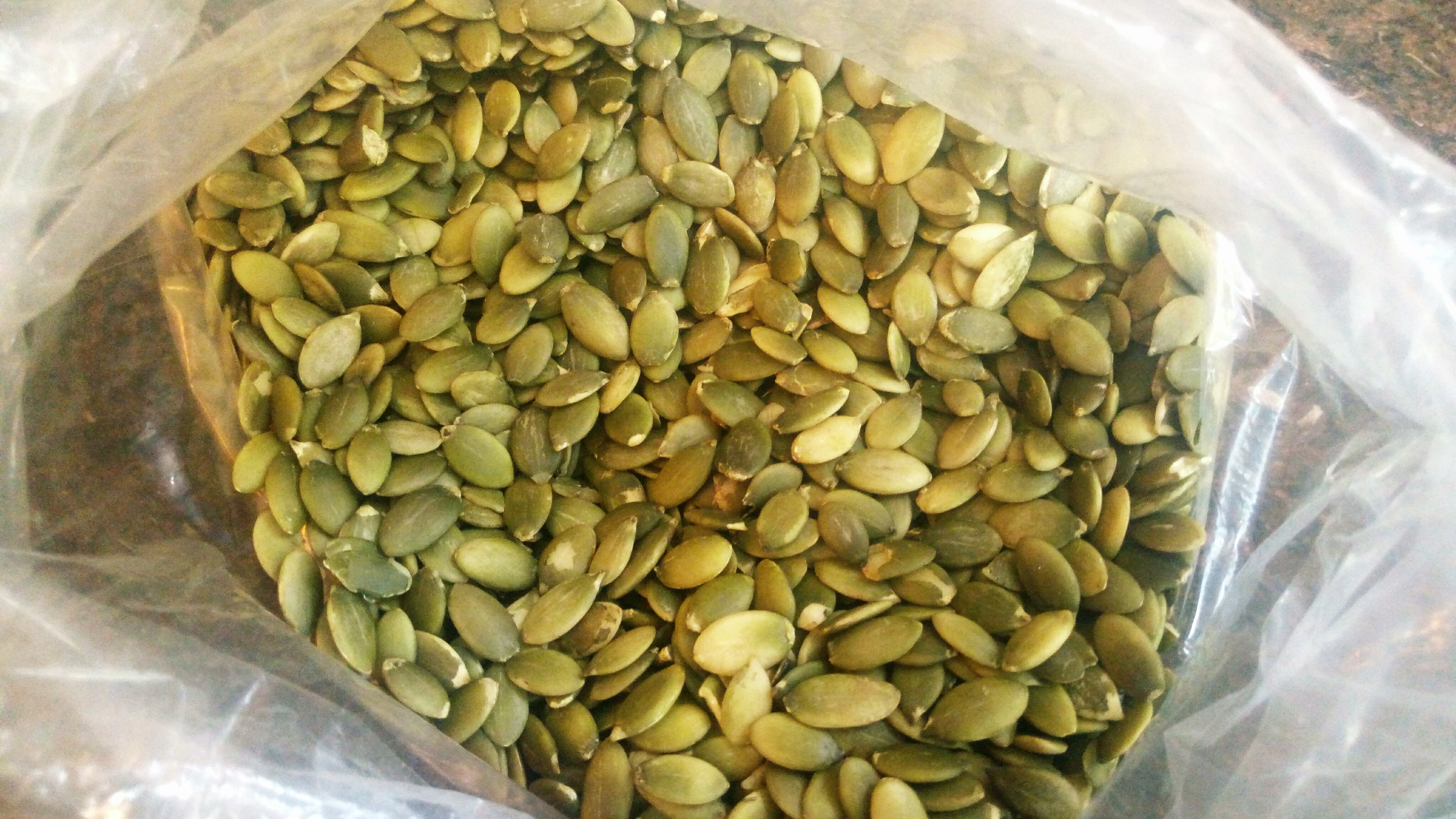
Pumpkin seed kernels, raw, unsalted.

Pumpkin seed kernels are a good source of protein, essential fats, fiber, magnesium and other vitamins and minerals – and a great source of zinc which may be lacking in vegetarian or vegan diets. (Pumpkin Seeds – Benefits, nutrition and dietary tips.) (Other vegetarian sources of zinc.)

Work is progressing on the development of pumpkin seed flour for use as a food thickening substance for use in gravies or other sauces or stews. It would increase the protein, essential fatty acids, and other trace nutrient content of the resulting foods. (10) The use of pumpkin seeds in the diet may also prove to be protective against cancer and liver or kidney injury; and as a good source of antioxidants such as carotenoids (vitamin A family of nutrients) the use of pumpkin seeds in the diet may prove to be helpful against many conditions that involve excess oxidative stress. (4, 5, 6, 7, 8, 9) They are also a source of vitamin E (tocopherols), other phytosterols, and linoleic acid, a beneficial polyunsaturated fat. Pumpkin seed oil may be helpful in wound healing. (15) Pumpkin seeds are also a good source of phospholipids, (16), which are important for skin and membrane health.

Pumpkins are considered a drought tolerant plant for gardeners. Adequate water is needed to grow larger pumpkin and squash but the vines can survive limited water conditions. The seeds of other summer and winter squash are also nutrient rich and also may be more drought tolerant plants. (11, 12) Enough but not too much water at the right times are critical. Flooding or severe drought may both harm the garden yield. Mulching and drip irrigation or other watering methods applied at optimal stages of growth can be the water thrifty solution for best yield. (13, 14)

The seeds of butternut squash and some types of winter squash can be collected when trimming the squash and later toasted and eaten as a crunchy nutritious snack. India grocery markets may also have shelled squash seeds available for sale. They are slightly smaller and paler in color than the shelled pumpkin kernels in the image above.

Magnesium is one of the beneficial nutrients found in pumpkin seeds. It is a mineral that is needed in greater quantity during pregnancy and high blood pressure/hypertension can be a symptom of deficiency. Preeclampsia and the more severe eclampsia are complications characterized by high blood pressure and edema/swelling that can occur during pregnancy. Toxins collect in the excess fluid buildup and can risk a brain condition similar to hypertension encephalopathy in the more severe eclampsia. Seizure activity can result and death for the woman and expected infant are risks. Magnesium is used as an intravenous or intramuscular injection to reduce risk of the seizures during eclampsia. The mineral seems to help protect the blood brain barrier and reduce swelling in the brain during eclampsia. It’s role as an antioxidant to reduce free radical toxins may be involved but the exact mechanism for its benefit in eclampsia is not known. (2, page 139)

More information about preclampsia is available in a previous post, and more information about food sources and supplement sources of magnesium is also available in a previous post.

I have several writing projects in draft mode however they overlap – pumpkin seeds, a good source of magnesium and zinc, helped my previous prenatal clients who had a history of preeclampsia or high blood pressure in a previous pregnancy prevent a reocurrence of the problem. Why? Possibly because of the magnesium and other beneficial nutrients which could be protecting the blood brain barrier and might then also be helpful for preventing harm to oligodendrocytes and protect against demyelination – a risk that can occur with some types of encephalopathy (3).

Disclaimer: Opinions are my own and the information is provided for educational purposes within the guidelines of fair use. While I am a Registered Dietitian this information is not intended to provide individual health guidance. Please see a health professional for individual health care purposes.

  1. https://www.ncbi.nlm.nih.gov/pmc/articles/PMC4827242/Megan Ware, What are the health benefits of pumpkin seeds?, July 24, 2018, https://www.medicalnewstoday.com/articles/303864.php
  2. Mehmet Kaya, Bulent Ahishali, Chapter 9: The role of magnesium in edema and blood brain barrier disruption, page 139, in the book edited by Robert Vink, Mihai Nechifor, Magnesium in the Central Nervous System, University of Adelaide Press, 2011, adelaide.edu.au, free ebook pdf, https://www.adelaide.edu.au/press/titles/magnesium/magnesium-ebook.pdf  (2
  3. S. Love, Demyelinating Diseases, J Clin Pathol. 2006 Nov; 59(11): 1151–1159.  https://www.ncbi.nlm.nih.gov/pmc/articles/PMC1860500/ (3)
  4. Scientific Studies collection on a commercial website by Pepo Farms, https://pepofarms.com.au/scientificstudies/ (4) which includes:
  5. M. Gossell-Williams, A. Davis, N. O’Connor, Inhibition of Testosterone-Induced Hyperplasia of the Prostate of Sprague-Dawley Rats by Pumpkin Seed Oil. Jun 2006, Vol. 9, No. 2 : 284 -286. 
  6. C. Z. Nkosi, A. R. Opoku, S. E. Terblanche, Antioxidative effects of pumpkin seed (Cucurbita pepo) protein isolate in CCl4-Induced liver injury in low-protein fed rats.
  7. Fahim AT Abd-el Fattah AA Agha AM Gad MZ
    Effect of pumpkin-seed oil on the level of free radical scavengers induced during adjuvant-arthritis in rats.
    In: Pharmacol Res (1995 Jan) 31(1):73-9 ISSN: 1043-6618
  8. Suphakarn VS Yarnnon C Ngunboonsri P, The effect of pumpkin seeds on oxalcrystalluria and urinary compositions of children in hyperendemic area. In: Am J Clin Nutr (1987 Jan) 45(1):115-21 ISSN: 0002-9165
  9. Matus Z Molnar P Szabo LG [Main carotenoids in pressed seeds (Cucurbitae semen) of oil pumpkin (Cucurbita pepo convar. pepo var. styriaca)] Olajtok (Cucurbita pepo convar. pepo var. styriaca) magjabol nyert presmaradek ossz-karotinoid-tartalmanak es karotinoid-osszetetelenek meghatarozasa. In: Acta Pharm Hung (1993 Sep) 63(5):247-56 ISSN: 0001-6659 (Published in Hungarian)  * The main carotenoids included per the Pepo Farms site: “The main components of the press-residue were lutein [3,3′-dihydroxy-alpha-carotene = (3R,3’R,6’R)-beta,epsilon-carotene-3,3′-diol; 52.5%] and beta- carotene (beta,epsilon-carotene; 10.1%). In addition to the above- mentioned pigments it was successful to reveal the presence of violaxanthin, luteoxanthin, auroxanthin epimers, lutein epoxide, flavoxanthin, chrysanthemaxanthin, 9(9′)-cis-lutein, 13(13′)-cis- lutein, 15-cis-lutein (central-cis)-lutein, alpha-cryptoxanthin, beta- cryptoxanthin and alpha-carotene (beta,epsilon-carotene) in small quantities.”  (4)
  10. Initial food technology research on the preparation and use of pumpkin seed flour for use in more nutritious gravy type sauces: Sharma G, Lakhawat S., Development, Quality Evaluation and Acceptability of Pumpkin Seed Flour Incorporated in Gravy. J Nutr Food Sci 7:613. doi: 10.4172/2155-9600.1000613      https://www.omicsonline.org/open-access/development-quality-evaluation-and-acceptability-of-pumpkin-seed-flourincorporated-in-gravy-2155-9600-1000613.php?aid=91345
  11. EllenB, Growing Drought Tolerant Vegetables, June 9, 2009, ThriftyFun.com, https://www.thriftyfun.com/Growing-Drought-Tolerant-Vegetables.html (11)
  12. Troy Scott, Drought Tolerant Vegetables for your Garden, July 9 2018, HeavenlyGreens.com http://www.heavenlygreens.com/blog/drought-tolerant-vegetables-for-your-garden (12)
  13. Joan Morris, Vegetable Gardening in a Drought, mercurynews.com, April 1, 2015,  https://www.mercurynews.com/2015/04/01/vegetable-gardening-in-a-drought/ (13)
  14. Extension Utah State University, Vegetable Irrigation: Squash and Pumpkin, Horticulture/Vegetables/2015-4,   https://digitalcommons.usu.edu/cgi/viewcontent.cgi?article=1744&context=extension_curall (14)
  15. Bardaa S, Ben Halima N, Aloui F, et al. Oil from pumpkin (Cucurbita pepo L.) seeds: evaluation of its functional properties on wound healing in rats. Lipids in Health and Disease. 2016;15:73. doi:10.1186/s12944-016-0237-0. https://www.ncbi.nlm.nih.gov/pmc/articles/PMC4827242/ (15)
  16. Zh.Y. Petkova, G.A. Antova, Changes in the composition of pumpkin seeds (Cucurbita moschata) during development and maturation. Grassas Y Aceites, 66 (1), Jan–March 2015, e058. http://grasasyaceites.revistas.csic.es/index.php/grasasyaceites/article/viewFile/1523/1658 (16)